
Tree of Life plasmahydroglycerol – Bark and Gum Resin
The “Tree of Life” Bark and Gum Resin are very healing and regenerating. The Bark and Gum Resin from the “Tree of Life” produce a byproduct, I named Plasmahydroglycerol Pro.
A few images have a defect which is noticeable, it was from a digital camera I purchased and had to return because of this camera defect. I apologize but the few that have it, I needed to share the entire image details and would not crop it out.
Ninety-seven percent (97%) of all micrographs of Plasmahydroglycerol and Gum resin were captured on digital camera inserted into ocular lens of my novel microscope using a 10x wide field and set at 4x object lens equaling 40x magnification. The remaining 3% of some Gum resin images were observed using a 10x wide field lens with a 100x objective lens equaling a 1000 x magnification. See here The “Tree of Life” rare and non-conventional scientific analytical evaluations.
Image Descriptions
Below images reveal Immortal Genetic prismatic molecules of the X-Chromosome with Centromere at junction and Telomeres at the ends of the long and short arms and Centrosomes with Centriole nucleus of Neutron Royal Purple Crosses, right angles, and a few square structures. Additionally, cytoskeletal cells, neurons, neuron cell bodies, Intermediate and Long filaments, myelin sheathing and axons, as well as dendritic neurons, Pyramidal neurons, Purkinje neurons, and Hematopoietic Stem Cells

Image 001-Liquid Plasmahydrogycerol-at 40x magnification shows an Evolutionary Single Chromatid comprised of adjacent elliptical concentric Centromeres with Royal Purple nucleus- on the right a larger Centromere of concentric rings-
Image 01-Liquid Plasmahydroglycerol-40x magnification-shows God’s genetic disk-shaped Mitotic Tubules converted to Centrosomes with a Royal Purple Centriole nucleus – looks like lunar eclipse


Image 02-Liquid Plasmahydroglycerol- Shows an Evolutionary Luminescent X-Chromosome in metastate with two connecting sister chromatids at the centromere junction and telomeres at the end of short arms pointing towards Nadir or South-1 .jpg[Signifying God’s short stay here on earth- his long arms point North where he stays in his heavenly Kingdom] Below is a schematic drawing of an X-Chromosome created in a Petri Dish or Invitro cloning- click on the link below to view in larger png file.
By File:Chromosome-upright.png Original version: Magnus Manske, this version with upright chromosome: User:Dietzel65 Vector: derivative work Tryphon – Own work based on: Chromosome-upright.png, CC BY-SA 3.0,
Diagram of a replicated and condensed metaphase eukaryotic chromosome: Chromatid Centromere Short arm Long arm
File:Chromosome-upright.png Original version: Magnus Manske, this version with upright chromosome: User:Dietzel65 Vector: derivative work Tryphon – Own work based on: Chromosome-upright.png
Scheme of a Chromosome. (1) Chromatid. One of the two identical parts of the chromosome after S phase. (2) Centromere. The point where the two chromatids touch, and where the microtubules attach. (3) Short (p) arm (4) Long (q) arm. In accordance with the display rules in Cytogenetics, the short arm is on top.


Image 2a -Myelinated_neuron- The Tree of Life is similar but far more STABLE-ELASTIC and REGENERATIVE.jpg
Image 2b-Axolemma_Histology, _OpenStax_College-Myelin Sheath and Axons

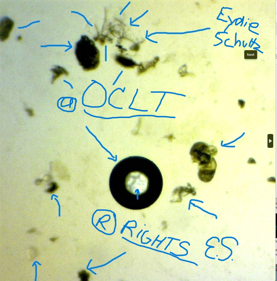
Image 2c- A Screenshot of Expanded view from Image 2d- shows a perfect spherical Myelin sheathing and Myelinated axon neurons
Image-2d- Liquid plasmahydro glycerol-40x magnification- shows a perfect spherical collagen rich Myelin sheathing and Myelinated axon neurons-


Image 2e-Liquid Plasmahydroglycerol-showing a single cytosol cluster of axons and glial cell at bottom base in center by blue arrow
Image 2f- gum resin 40x magnification shows God’s genetic long GNA single strand in loop crossing position-1.jpg – [God is not composed of DNA nucleotide poisons-So I cannot use DNA in any description of images


Image 3– Liquid plasmahydroglycerol- 40x magnification shows a neural network of short dendritic neurons-axons and Nissl bodies
Image 4-Gum resin shows Fibrous Microtubule Green tracts on left just below compact quaternary structure of chromatids and above-Golgi apparatus is near bottom center and directly above is mitotic spindle poles centrosomes and centrioles


Image 5-gum resin at 400x magnification shows Primitive Plasma membrane nucleus with genetic fragments-lysosome autophagy invagination at top center on the left-a few flat circular vesicles-axon neurons-many glial cells and glial formation in center image-1 .jpg [I placed arrows by myelin sheathing in this image.]
Image 6-gum resin 400x magnification shows membrane nucleus-Golgi apparatus circled-Centrosomes and Centriole blue circled-myelin sheaths-circular vesicles- dendrite neurons- interneurons- microglial cells-Mitotic Tubules-green fiber tracts


Image 6a Cytokinesis-electron-micrograph.jpg centrosome at arrow and nuclei.
Image 7-gum resin 40x magnification-shows Evolutionary cell body of a neuron with a transparent nuclear envelope a nucleolus with compacted chromatids-copper fibrous mitochondria and transparent dendritic axons on surface-


Image 8- Expanded Human cell Nucleus 2024-02-01 164013- invitro schematic drawing- similar to image 7 of God’s Neuron cell-1.jpg.
By BruceBlaus – Own work, CC BY-SA 4.0, https://commons.wikimedia.org/w/index.php?curid=46621398
Image 8B diagram of neuron components
https://en.wikipedia.org/wiki/Neuron#/media/File:Blausen_0657_MultipolarNeuron.png


Image 8c- Schematic of an anatomically accurate single pyramidal neuron, the primary excitatory neuron of the cerebral cortex, with a synaptic connection from an incoming axon onto a dendritic spine
By Amyleesterling – Own work, CC BY-SA 4.0, https://commons.wikimedia.org/w/index.php?curid=65952135
This schematic shows a single pyramidal neuron, the dominant excitatory neuron of cerebral cortex, with an synaptic connection from an incoming axon. It briefly explains the soma, dendrites, axon, and synapse and was created by Amy Sterling and Daniela Gamba for Neo Brain Game.
- CC BY-SA 4.0
- File:Anatomy of a Neuron with Synapse.png
- Created: 28 January 2018
- Uploaded: 29 January 2018
https://en.wikipedia.org/wiki/Neuron#/media/File:Anatomy_of_a_Neuron_with_Synapse.png
Image 9-Gum resin 40x magnification-shows Primitive inner Plasma membrane neuron cell with embedded monocytes in transparent axons and dendrite branching and on surface- condensed fibrous chromatids in nuclei and within X-Shaped copper mitochondria-1.jpg. [Upon Expanding this image, it is evident of the monocytes attached to transparent axons and dendrite branching]


Image 10-Gum resin 40x magnification-shows Primitive inner Plasma membrane neuron cell with embedded monocytes in transparent dendrite branching and on surface- condensed fibrous chromatids in nuclei and within X-Shaped copper mitochondria-1.jpg.
Image 11-Gum resin-40x magnification-shows Primitive brain neuron with embedded monocytes within transparent axons and dendrite branching- Cisternae and trans at center bottom-Golgi apparatus circled in blue- and mitochondria-1.jpg. [It seems as if the monocytes are a conversion to some neurons because they are anchored at the root or formation. Upon expansion this image shows Purkinje neurons and cells]


Image12 –Gum resin-40x magnification-shows unipolar transparent axon dendrite neuron with enveloped monocytes between inner and outer gate opening-and formation of monocyte within blue circle-1.jpg.
Image-12a-Gum resin-40x magnification-shows inner membrane of Primitive neuron with monocytes by all blue marked arrows-a few Nissl bodies- Circular vesicles in early formation of isolated fibrous midtone colorful microtubule centrosomes-1.jpg


Image13a-Gum resin-shows monocytes enveloped in cortical surface and brain afferent sensory neurons- a few black fibrous bulbous bodies anchored at ends of transparent monocyte dendrite branching-1 .jpg [In my opinion, it’s as if the stem cells form and can even convert into neurons]
Image13– Gum resin-40x magnification- shows cluster of flat sensory interneurons with attached monocytes anchored to the smooth cortical surface-and monocytes enveloped in long axon dendrites and thick Purkinje neurons with flat fibrous bulbous-1.jpg.


Image14a Purkinje Cell or neurons- to compare with the Tree of life brain cells in Image 13.jpg.
Image14b -Cerebellum_-_biel_-_very_high_mag-Purkinje cells- to compare to Tree of Life brain cells in a few other of my micrographs or images .jpg


Image14-gum resin-at 40x magnification-shows a few unipolar afferent or sensory neurons with a flat bulb at one end- a few interneurons-Nissl bodies and singular monocytes anchored to cortical surface-1.jpg.
Image15– Gum resin-image 40x magnification shows anchored Purkinje neurons-and Purkinje gold cells-long unipolar sensory neuron with enveloped monocytes and flat bulb-isolated monocytes by blue arrows and neural stem cells by gate opening-1.jpg.


Image 16: Gum resin-40x magnification-shows a few early developing Purkinje neurons with enveloped monocytes-anchored to cortical smooth surface-similar to a single pyramid neuron-and one circled glial cell- a few isolated monocytes
Image16a -Anatomy_of_a_Neuron_with_Synapse- Schematic drawing of a Single Pyramid Neuron-from bioengineered protocols is similar to Image 16– God’s early developing Purkinje neurons from the micrographs of the Tree of Life-1.jpg [The early developing Purkinje neurons with enveloped monocytes in Image 16 do resemble the Single Pyramid neuron in the study of neuroscience schematic drawing from in-vitro bioengineered protocols.]


Image17-Gum resin 40x magnification shows two isolated singular Purkinje neurons blue circled and near upper right corner next to small gold colored Purkinje cells all anchored along with monocytes to smooth cortical surface-1.jpg.
Image18: Liquid plasmahydroglycerol-shows Primitive Evolutionary dense porous cortical bone surface with an osteoblast of enlarged irregular shaped elastic collagen rich membrane- and a few basophiles anchored to surface-1.jpg.
https://commons.wikimedia.org/wiki/File:Bonemetabolism.svg#/media/File:Bonemetabolism.svg
Bone tissue is removed by osteoclasts, and then new bone tissue is formed by osteoblasts. Both processes utilize cytokine (TGF-β, IGF) signaling.
TGF-B- Transforming Growth Factor- Beta
IGF-Insulin Growth Factor
Ossification Bone metabolism
[However, GOD’S is instantaneous with Structural Regulation of Division and Growth always retaining Homeostasis]


Image19-Liquid Plasmohydroglycerol-shows-blue marked Evolutionary dense porous cortical surface with one enlarged and one smaller irregular shaped osteoblast elastic collagen rich membrane and many anchored basophils on bone surface and gold marrow-1.jpg
Image20-Liquid Plasmahydroglycerol- shows Evolutionary white matter cortical smooth bone surface with small divets-1 .jpg


Image21-Liquid Plasmahydroglycerol-shows a small round elastic collagen rich osteoclast membrane anchored near anchored basophile attached at base surface to compact osteoclast performing apoptosis on dense semi-porous cortical bone surface-1.jpg.
Image22-liquid plasmahydroglycerol-shows collagen rich irregular osteoblast and round osteoclasts anchored to a smooth slightly porous cortical bone surface with monocyte neural stem cells by blue arrows-1.jpg.


Image23-Gum resin- 40x magnification shows copper gold collagen rich barbed end intermediate filaments or fibroblast-many Prismatic Genetic Centrosomes with Centriole Royal Purple nucleus-1.jpg [Upon expanding this image many Prismatic Genetic Centrosomes with Centriole Royal Purple nucleus showing interstate-prostate-anastate and telostate mitotic division and replication]
Image24– Gum resin- shows copper rich collagen barbed and pointed end intermediate filaments and many Prismatic early developing genetic centromeres converting to centrosome and centriole formations-1.jpg. [The royal purple molecular structure of metastate Crosses, right angles, and square patterns of reflection-rotation-translation derive from 2D dimensional elliptical Centromeres with concentric rings. So, God’s Genetic molecules also differentiate or convert from the Centromeres to Centrosome and Centrioles (not Centrosomes of fibrous coiled strands, but of prismatic Centromeres originating from a metastate X-Chromosome as shown in Image-02. There are Fibrous Mitotic Green Tubules, Centrosomes and Centrioles in Images 4 and 6 and other images with fibrous chromatids. I think this represents Jesus when he walked here on earth in half man and half immortal physique,]


Image25-Gum resin-40x magnification-shows copper rich collagen intermediate and microfilament Actins with barbed and pointed ends-1.jpg.
Image26-Gum resin 40x magnification shows copper collagen rich oblate microfilaments with extending lateral pointed ends-large spatial regions with Mitotic Green Tubules in various shapes-some elliptical Centromeres with concentric rings-1.jpg [Note: blue arrows highlight formation of Green Microtubules behind the Prismatic Centrosomes]


Image27 Gum resin-40x magnification-shows Prismatic prolate and elliptical Genetic Centromeres with concentric ring-copper rich collagen microfilaments and early Mitotic Spindle formation circled by blue arrows-1.jpg.
Image 28: Gum resin- 40x magnification showing copper rich collagen Microfilaments-Intermediate Filaments-Prismatic Genetic Centromeres- converting to early formation Mitotic Spindles-some Centrosomes with Royal Purple Centriole nucleus-


Image28a MEF_microfilaments- Microfilament (actin cytoskeleton) of mouse embryo fibroblasts, stained with FITC-phalloidin (100-fold magnification.)- some comparison to God’s Intermediate Filaments .jpg
Image28b: Cytoskeletal_Components- Eukaryte cytoskeleton-tubulin dimers-similar but different than God’s Centrosome and Centriole .jpg [The tubulin dimers are different in shape and fibrous structure in comparison to God’s Microtubule highly organized spherical structure and centriole with a molecular Neutron Cross].
Image 28bThe Cytoskeleton consist of a. microtubules b. microfilaments c. intermediate filaments.
Author attributes
By OpenStax – https://cnx.org/contents/FPtK1zmh@8.25:fEI3C8Ot@10/Preface, CC BY 4.0, https://commons.wikimedia.org/w/index.php?curid=30131202

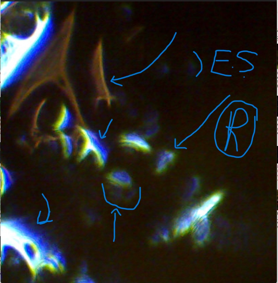
Image28c Gum resin-A Screenshot expansion of Image28 showing early states of Genetic Centromeres converting to Mitotic spindle strands and early formations of Centrosomes and Centrioles with Royal Purple nucleus-1 .jpg
Image29 Liquid Plasmahydroglycerol-40x magnification shows God’s Genetic Prismatic Mitotic Spindle nucleating into amorphous condensed optical fibers with some Centrosomes and early states of Royal Purple Centrioles-1.jpg


Image30- Liquid Plasmahydroglycerol -40x magnification-Upon expansion shows Mitotic Tubules converted to Luminous Centrosomes and Centrioles-within the Alpha Super Helical GNA and on the right side-1 .jpg [note- The Centrosomes and Centrioles with Royal Purle nucleus and neutron molecular structures are formed in a variety of shapes embedded in the Alpha Super Helical GNA. Additionally, on the right side of the Alpha Super Helical GNA shows different states of mitotic division and replication of Metastate, Prostate, Anastate and Interstate .jpg
Image31-Gum resin-40x magnification- shows a population of Evolutionary Genetic Confined Centrosomes and early state formation of Royal Purple Centrioles with macromolecular neutrons at interstate, metastate, anastate and prostate-1 .jpg [Centromeres and Telomeres at the terminal of prismatic metastate X-chromosome as shown in Image-02 coverts to Centrosomes and Centrioles as shown in Image-24, Image 26, Image 30-32 expansion, Image 28c, Image 29 and other images.


Image32-Gum resin- A Screenshot Expansion of Image 31 shows a population of Evolutionary Confined Centrosomes and Centrioles in mitotic division and replication of Genetic Royal Purple macromolecular neutron structures
Image33-Liquid Plasmahydrogycerol-shows double stacked Centromeres and singular Centromeres-1.jpg. [The Evolutionary Prismatic centromeres are the junction of the Prismatic X-Chromosome or two sister chromatids- as shown in Image02 an in metastate.


Image34-Liquid Plasmahydro gycerol- shows two Large Evolutionary Prismatic Centromeres near bottom right corner- expanding this image shows prismatic production of various Centrosomes and Centrioles with Royal Purple molecular structures of Crosses, straight lines, and right angles spatially apart from one another showing mitotic division Interstate-Anastate-Metastate and Telostate .jpg
[https://link.springer.com/article/10.1007/s41048-019-00101-x]
https://link.springer.com/article/10.1007/s41048-019-00101-x
images and information about bioengineered Centrosomes and Centrioles]
Image-35-Image-35-Liquid-Plasmahydro glycerol-shows Evolutionary luminescent flat disk and some elongated nucleated Centrosomes with Centrioles of many Royal Purple Neutron macromolecular structures of Crosses, right angles- orthogonal and square patterns .jpg [This image also shows the head of a SEAHORSE in vivid colors]


Image-36 Liquid plasmahydroglucerol-40x magnification-A blue marked and highlighted version of Image 35 to show God’s luminescent Genetic disk and elongated Centrosomes-with Centrioles of many Royal Purple CROSSES- Right Angles-geometric patterns and a seahorse head
Image-37-Liquid plasmahydroglycerol-40x magnification- A Screenshot Expanded view from Image 35 and Image 36 blue marked Evolutionary Centrosomes and Centrioles with macromolecular Neutron Royal Purple CROSSES and a SEAHORSE HEAD .jpg [This image magnifies the Centrioles Neutron Macromolecular Neutron Structures of Royal Purple Crosses and Reflective Background- Orthogonal or Right Angles. The Centrosomes are spatially apart with yellow/orange/red/green disk and spherical nucleated shapes in mitotic division and replication. The Centrosome on the right show more Green/Blue colors and the Centriole shows a Royal Purple Cross with a Reflective Background of faded Cross.]


Image-37a- Liquid Plasmahydroglycerol- 40x magnification-A Screenshot Expanded view from Image 35 and 36 showing two centrosomes near the bottom The one on the left is in Anastate with separate centrioles at right angle rotations-The one on the right shows a neutron macromolecular structure of a Cross
Image-38 –Liquid Plasmahydroglycerol- 40x magnification-A Screenshot expansion from Image-35 showing many Centrosomes and Centrioles with Royal Purple neutron molecular CROSSES-2 .jpg
[Also, Royal Purple neutron molecular structures at right angles, faded Cross in reflection, rotation, and translations and one square pattern. All Centrioles show mitotic division in Interstate, Anastate, Metastate, and Telostate]


Image-39-Expansion view from Image 35-36 and 38 shows Evolutionary GNA Genetic Centrosomes and Centriole nucleus with Royal Purple Neutron macromolecules of a Cross and right angles in Telostate and Anastate on left and metastate on right
Image-39a Bioengineered through in vivo and in vitro processes- a schematic drawing of Laminin111 molecular cross structure- doesn’t even come close to God’s Genetic macromolecules micrograph or images from the Tree of Life CROSSES


Image-40 A Screenshot Expansion from Image-38- Shows Evolutionary Genetic disk and elongated shaped Centrosomes with Centrioles in Royal Purple Neutron macromolecular structures of Crosses and right angles in reflection-rotation and translation
Image-41– Image-41- A Screenshot Expansion from Image-38-God’s Evolutionary Centrosomes and Centriole Genetics of luminescent Royal Purple Neutron macromolecular structures of faded CROSS on left in metastate- square pattern of reflecdtion at center in Prometastate- and right-angle reflections on right in Telostate


Image-42– Image-42-Liquid Plasmahydrogycerol- at 40x magnification shows vivid Prismatic Telomeres from the short arm ends of the Evolutionary X-Chromosome micrograph from Image-02
Image-43-Liquid Plasmahydroglycerol- shows neural network of white amorphous fiber optic pyramidal neurons-blue circle shows defect from a defective digital camera- my Initials- to mark my property rights on all micrograph images-1 .jpg [This image resembles Image 45]


Image-44-Liquid Plasmahydroglycerol showing neural network of white amorphous optic fiber pyramidal neurons- blue marked showing defect from camera-and tiny centrosomes-1 .jpg [I had to return the new digital camera I purchased because of this blue marked defect that is seen in many images. The pyramidal neurons from the Tree of Life do not show a bulbous protrusion, but it does have many flat larger nodes- also this image shows beautiful tiny centrosomes near top by camera defect- Pyramidal Neurons are in the hippocampus area within the cerebral cortex region]
Image-45-Liquid Plasmahydroglycerol- 40x magnification shows white fiber optic dendritic strands and isolated pyramidal spiny neurons with flat nodes and near camera defect shows larger centrioles with royal purple neutron macromolecules


Image-46-Z_MaxProjection_of_MediumSpinyNeurons_Gpr101Cre_dtTomato-from mouse substantia nigra- a red fluorescent protein used.jpg [There are similarities of the mouse’s pyramidal spiny neurons, but still different from God’s Pyramidal Neurons.]
Image-46– Liquid Plasmahydro glycerol- enlarged view of a single white fiber optic Pyramidal Neuron- with a large flat bulbous -faded and blurry Royal Purple prisms in the loop- and dendritic branching at top near end terminal


Image-46a– Gyrus_Dentatus_40x Golgi-stained neurons in human hippocampal tissue .jpg https://commons.wikimedia.org/wiki/File:Gyrus_Dentatus_40x.jpg#/media/File:Gyrus_Dentatus_40x.jpg
The link above shows Golgi-stained neurons in human hippocampal tissue.
By Methoxy Roxy – Own work, CC BY-SA 2.5, https://commons.wikimedia.org/w/index.php?curid=1325252
https://en.wikipedia.org/wiki/Neuron#/media/File:Smi32neuron.jpg
This above link also shows stained Pyramidal neurons in cerebral cortex.
Image-47- Liquid Plasmahydroglycerol-enlarged view of a white amorphous single pyramidal neuron- with a large flat bulbous near tail end and a royal purple prisms glassy loop strand attached to flat bulb and at opposite terminal end dendrite branch
